PET scan
A PET scan is an examination that provides information about the metabolism in the body. It is a nuclear test in which radioactive nutrients (for example, sugars and proteins) are administered to a patient. In places with an increased metabolism, such as in a malignant tumor, more nutrients are absorbed. This can be visualized with a PET camera.
The device on which PET scans are made is very similar to that of a CT scan (see image)
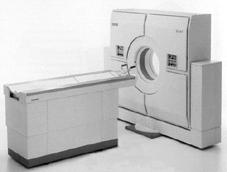
Before the patient lies on the table, the liquid containing radioactive substances is injected. It must first spread through the body.
A PET scan is not a standard examination for patients with liver disease. If a CT or MRI scan is inconclusive, patients with colorectal cancer suspected of metastasis (including to the liver) are eligible for a PET scan.
The Erasmus Medical Center does not yet have a PET scan. Patients are sent to Antwerp or Nijmegen for the scan, after which the results are discussed here.
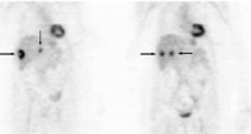
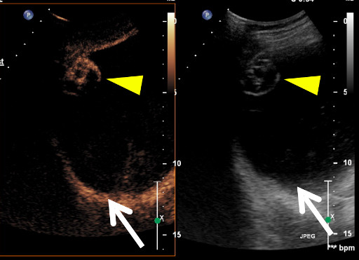
These scans show a longitudinal cross-section through the body. The dark spots indicated by the arrows are areas of increased radioactivity located in the liver. These indicate the presence of metastases in the liver.
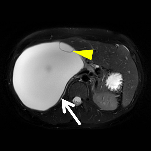
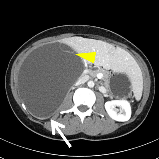
An ultrasound, CT scan and an MRI examination provide other information about the nature of the abnormality. Depending on the nature of the abnormality, a combination of tests will be required to make a proper diagnosis.