CT scan
The CT scan (CT = Computer Tomography) uses X-rays. X-rays were discovered in 1895 by Professor Wilhelm Roentgen and have the property that they can penetrate through the body and are absorbed to different extents by the different tissues in the body. Subsequently, the rays that have passed through the body are absorbed by a film, after which a photo can be developed. With this, an imaging technique was developed that the CT scan also uses.
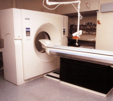
A CT scan is a round device into which the patient is inserted. A beam projector and detector circle the patient, and 'slices' are then made of the body. The radiologist looks at these pictures and assesses any abnormalities that are found. The examination takes about 20 minutes, and the patient does not feel anything. Several series of photos are taken during the investigation.
In some cases, a CT scan is not sufficient to distinguish between different conditions and an MRI scan can be made.
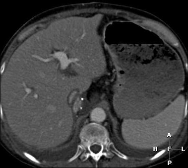
What you see is a cross section through the body. The bottom of the picture is the spine and you see a vertebra. The top is the belly side. You have to imagine looking at the cut body from below and then the left side of the picture is the right side of the body. The large organ that can now be seen on the left of the picture is the liver. The white structure in the center is one of the major blood vessels of the liver.