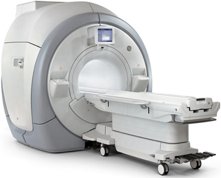
MRI scan
An MRI scan (MRI = Magnetic Resonance Imaging) uses magnetic energy and radio waves to make cross-sections, or slices, of the body. An MRI scan consists of a tube and a cylinder-shaped magnet.
During the examination, a signal is emitted, the energy of which is partly absorbed by the atoms of the body and partly reflected back. These reflected signals are measured by a computer and converted into an image. An advantage of the MRI examination is that the MRI scan can create images in all directions, and not just transverse slices. Making an MRI scan may be necessary to distinguish between various abnormalities that cannot be distinguished with a CT scan.
For the examination, the patient is placed in a kind of tube, which is quite narrow. The examination lasts approximately 40 minutes, and several series of photos are taken during the examination. The patient feels nothing further from the examination.
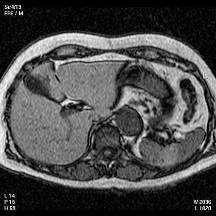
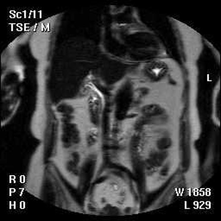
Below is an example of an MRI scan of a normal liver in two different directions.